Earlier research in biomedical image reconstructions
includes
brain reconstructions from
compressed sensing
and 3D cardiac reconstructions
from freehand ultrasound images.
Also, for general images,
refer to the stochastic methods described in
“Multiscale Sampling Geometries
and Methods for Deterministic and Stochastic
Reconstructions of Magnitude and Phase Spectra
of Satellite Imagery”.
Summaries and perspectives on research from different groups
can be found in editorials in special issues
and the telemedicine review papers.
For commercial applications, refer to
the ivPCL commercialization webpage.
You can also share website information.
Recent papers
-
Panayides, A., Pattichis, M.S., Leandrou, S., Pitris, C.,
Constantinidou, A., and Pattichis, C.S., “Radiogenomics for Precision
Medicine With A Big Data Analytics Perspective,” IEEE Journal of Biomedical
and Health Informatics, in press, 2019.
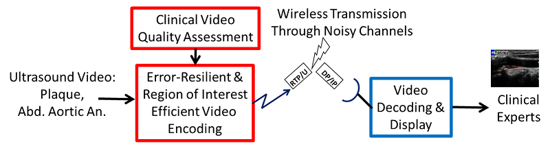
Medical ultrasound video communications
The goal of the project is to develop advanced methods for
communicating medical ultrasound video through noisy,
wireless channels without
compromizing diagnostic quality.
For effective communications, the developed systems need to
respond to bandwidth changes, video packet drops,
and user requirements for high frame rates, high resolution, and low delay.
- Panayides, A.S., Pattichis, M.S., Loizou, C.P., Pantziaris, M., Constantinides, A.G., and Pattichis, C.S.,
“
An Effective Ultrasound Video Communication System Using Despeckle Filtering and HEVC,”
IEEE Journal of Biomedical and Health Informatics,
vol. 19, no. 2, pp. 668-676, March 2015.
- Panayides, A., Pattichis, M.S., and Pattichis, C.S.,
“
M-Health Systems Use Diagnostically Driven Medical Video Technologies,” (invited),
IEEE Signal Processing Magazine,
vol. 30, no. 6, pp. 163-172, November 2013.
- Panayides, A., Antoniou, Z., Mylonas, Y., Pattichis, M.S., Pitsillides, A., Constantinides, A.G., Pattichis, C.S.,
“High-Resolution, Low-delay, and Error-resilient Medical Ultrasound Video Communication Using H.264/AVC Over Mobile WiMAX Networks,”
IEEE Journal of Biomedical and Health Informatics,
pp. 619-628, May 2013.
- Panayides, A., Pattichis, M.S., Pattichis, C.S., Loizou, C., and Pitsillides, A.,
“A Tutorial for Emerging Wireless Medical Video Transmission Systems,”
IEEE Antennas & Propagation Magazine,
vol. 53, no. 2, pp. 43-50, April 2011.
- Panayides, A., Pattichis, M.S., Pattichis, C.S., Loizou, C.P., Patziaris, M., and Pitsillides, A.,
“Atherosclerotic Plaque Ultrasound Video Encoding, Wireless Transmission, and Quality Assessment Using H.264,”
IEEE Transactions on Information Technology in Biomedicine,
vol. 15, no. 3, pp. 387-397, May 2011, PMID: 21233053.
Telemedicine
The goal of the telemedicine project is
to transmit biosignals, images, and videos anywhere, anytime
through the use of existing wireless networks.
Technological challenges arise from the need to
transmit larger datasets using limited bandwidth.
- Panayides, A., Pattichis, M.S., Pattichis, C.S., Loizou, C.,
and Pitsillides, A.,
“A Tutorial for Emerging Wireless Medical Video Transmission Systems,”
IEEE Antennas & Propagation Magazine,
vol. 53, no. 2, pp. 43-50, April 2011.
- Kyriacou, E., Pattichis, M.S., Pattichis, C.S., Panayides, A., and Pitsillides, A.,
“m-Health e-Emergency Systems: Current Status and Future Directions,”
invited in
IEEE Antennas and Propagation Magazine,
vol. 49., no. 1, pp. 216-231, Feb. 2007.
- Pattichis. C.S., Kyriacou, E., Voskarides, S., Pattichis, M.S., Istepanian, R., and Schizas, C.N.,
“Wireless Telemedicine Systems: An Overview,”
invited in
IEEE Antennas and Propagation Magazine,
vol. 44, no. 2, pp. 143-153, April 2002.
A historical video describing the EMERGENCY 112 project described above is
provided here by Professor C. Pattichis (an ivPCL affiliate and visitor).
Historic (low-resolution) video of Emergency 112 project.
Please do not maximize the window size unless viewing from a distance.
Stroke ultrasound image and video analysis
The goal of the stroke project is to
develop novel image processing methods that
can help assess the risk of stroke events as early as possible.
We have shown that early detection and risk assessment is indeed possible.
For emergency cases, refer to the
mobile video communications,
and telemedicine projects.
- Loizou, C.P., Murray, V., Pattichis, M.S., Pantziaris, M., Nicolaides, A.N., and Pattichis, C.S.,
“Despeckle
Filtering for Multiscale Amplitude-Modulation Frequency-Modulation (AM-FM) Texture
Analysis of Ultrasound Images of the Intima-Media Complex,”
International Journal of Biomedical Imaging,
vol. 2014 (2014), Article ID 518414, 13 pages.
- Kyriacou, E., Petroudi, S., Pattichis, C.S., Pattichis, M.S., Griffin, M., Kakkos, S., and Nicolaides, A.,
“Prediction of High Risk Asymptomatic Carotid Plaques Based on Ultrasonic Image Features,”
Special issue on “Atherosclerotic Cardiovascular Health Informatics Risk Screening and Intervention,”
IEEE Transactions on Information Technology in Biomedicine,
vol. 16, no. 5, pp. 966-973, 2012.
- Lambrou, A., Papadopoulos, H., Kyriacou, Pattichis, C.S., Pattichis, M.S., Gammerman, A., and Nicolaides, A.,
“Evaluation of the Risk of Stroke With Confidence Predictions Based on
Ultrasound Carotid Image Analysis,”
invited to Special Issue on Selected Papers from AIAI 2010,
International Journal on Artificial Intelligence Tools,
vol. 21, no. 4, 1240016, 18 pages, 2012.
- Sergio, M., Pattichis, M.S., and Barriga, E.S.,
“A Review of Motion Estimation Methods for Non-Invasive Ultrasound Motion
and Emerging Strain Imaging Methods of Carotid Artery Plaques,”
International Journal of Experimental and Computation Biomechanics,
vol. 1, no. 4, pp. 359-380, 2011.
- Loizou, C.P., Murray, V., Pattichis, M.S., Pantziaris, M., and Pattichis, C.S.,
“Multiscale Amplitude-Modulation Frequency-Modulation (AM-FM)
Analysis of Ultrasound Images of the Intima and Media Layers of the Carotid Artery,”
IEEE Transactions on Information Technology in Biomedicine,
vol. 15, no. 2, pp. 178-188, 2011, PMID 20889436.
- Kyriacou, E., Pattichis, C.S., Pattichis, M.S., Loizou, C.P., Christodoulou, C., Kakkos, S. and Nicolaides, A.,
“A Review of Non-invasive Ultrasound Image Processing Methods in the
Analysis of Carotid Plaque Morphology for the Assessment of Stroke,”
IEEE Transactions on Information Technology in Biomedicine,
vol. 14, no. 4, pp. 1027-1038, July 2010, PMID 20378477.
- Loizou, C.P., Pantziaris, M., Pattichis, M.S., Kyriakou, E. and Pattichis, C.S.,
“Ultrasound Image Texture Analysis of the Intima and
Media Layers of the Common Carotid Artery and its Correlation with Age and Gender,”
invited from conference paper presentation in IEEE BIBE 2008,
Computerized Medical Imaging and Graphics (Elsevier),
vol. 33, no. 4, pp. 317-324, June 2009, PMID: 19304453.
- Kyriacou, E., Pattichis, M.S., Pattichis, C.S., Mavrommatis, A., Christodoulou, C.I., Kakkos, S. and Nicolaides, A.,
“Classification of Atherosclerotic Carotid Plaques Using Morphological
Analysis on Ultrasound images,”
(a best conference paper award) invited in special issue on
Emerging Artificial Intelligence Applications and Innovations: Papers from AIAI 2006,
Journal of Applied Intelligence, Springer,
vol. 30, no. 1, pp. 3-23, February 2009.
- Kyriacou, E.C., Pattichis, C.S., Karaolis, M.A., Loizou, C.P., Christodoulou, C.I., Pattichis, M.S., Kakkos, S., and Nicolaides, A.,
“An Integrated System for Assessing Stroke Risk,”
IEEE Engineering in Medicine and Biology Magazine,
Special Issue on Image, Signal and Distributed Data Processing for Networked e-Health Applications,
vol. 26, no. 5, pp. 43-50, Sept.-Oct. 2007, PMID: 17941322.
Brain image analysis
A non-stationary image representation model for assessing multiple sclerosis cases
has also been applied here.
Please refer to the AM-FM representations projects for
more information on the approach.
- Loizou, C.P., Murray, V., Pattichis, M.S., Seimenis, I., Pantziaris, M., and Pattichis, C.S.,
“Multiscale Amplitude-Modulation Frequency-Modulation (AM-FM) Texture Analysis of
Multiple Sclerosis in Brain MRI Images,”
IEEE Transactions on Information Technology in Biomedicine,
vol. 15, no. 1, pp. 119-129, Jan. 2011, PMID 21062681.
Eye image analysis
The development of multiscale AM-FM representations
provided novel image features
that have been used in detecting eye diseases.
Please refer to the AM-FM
representations projects for
more information on the approach.
Earlier research focused on the development
of independent component analysis methods.
-
Agurto, C., Yu, H., Murray, V., Pattichis, M.S., Nemeh, S., Barriga, S., and Soliz, P.,
“A Multiscale Decomposition Approach to Detect Abnormal Vasculature in the Optic Disc,”
Computer Graphics and Image Processing,
vol. 42, pp. 137-149, July 2015.
-
Agurto, C., Murray, V., Yu, H., Wigdahl, J., Pattichis, M.S., Nemeth, S., Barriga, S., and Soliz, P.,
“A Multiscale Optimization Approach to Detect Exudates in the Macula,”
IEEE Journal of Biomedical and Health Informatics,
vol. 18, no. 4, pp. 1328-1336, 2014.
-
Yu, H., Barriga, E.S., Agurto, C., Echegaray, S., Pattichis, M.S., Bauman, W., and Soliz, P.,
“Fast Localization
and Segmentation of Optic Disc in Retinal Images
Using Directional Matched Filtering and Level Sets,”
IEEE Transactions on Information Technology in Biomedicine,
vol. 16, no. 4, pp. 644-657, July 2012.
-
Agurto, C., Barriga, S., Murray, V., Nemeth, S., Crammer, R., Bauman, W., Zamora, G., Pattichis, M.S., and Soliz, P.,
"Automatic Detection of Diabetic Retinopathy and Age-Related Macular Degeneration in
Digital Fundus Images,"
Investigative Ophthalmology and Visual Science,
vol. 52, no. 8, pp. 5862-5871, July 2011, PMID: 21666234.
-
Barriga, E.S., Pattichis, M.S., Ts’o, D., Abramoff, M., Kardon, R., Kwon, Y. and Soliz, P.,
“Independent Component Analysis using Prior Information for Signal
Detection in a New Functional Imaging Systems of the Retina,”
Medical Image Analysis,
vol. 15, no. 1, pp. 35-44, February 2011, PMID 20655800.
-
Agurto, C., Murray, V., Barriga, E., Murillo, S., Pattichis,
M.S., Davis, H., Russell, S.R., Abramoff, M.D., and Soliz, P.,
“Multiscale AM-FM Methods for Diabetic Retinopathy Lesion Detection,”
IEEE Transactions on Medical Imaging,
vol. 29, no. 2, pp. 502-512, February 2010, PMID: 20129850.
-
Barriga, E. S., Pattichis, M. S., Ts’o, D., Abramoff, M., Kardon, R., Kwon, Y., and Soliz, P.,
“Spatiotemporal independent component analysis for the detection of functional
responses in cat retinal images,”
IEEE Transactions on Medical Imaging,
vol. 26, pp. 1035-1045, Aug. 2007, PMID: 17695124.
Chest radiograph image analysis
The chest radiograph image analysis project
led to the development of a
computer aided diagnosis (CAD) system for detecting lung diseases.
The reader journal paper given below provides methods for
combining ratings from multiple experts.
- Pattichis, M.S., Soliz, P. and Cacoullos, T.,
“New Models for Region of Interest Reader Classification Analysis in Chest Radiographs,”
Pattern Recognition,
Special Issue of Digital Image Processing and Pattern Recognition Techniques for the Detection of Cancer,
(doi: 10.1016/j.patcog.2008.09.021),
vol. 42, no. 6, pp. 1058-1066, June 2009.
- Soliz, P., Pattichis C.S., Pattichis M.S., James D., and Ketai, L.,
“Texture analysis of opacity profusion in chest radiographs of miners with pneumoconiosis,”
American Journal of Respiratory & Critical Care Medicine, 2002; 165:A529
(short (< 1 page) journal publication of conference abstract). J65. Pattichis, M.S., Soliz, P., Pattichis, C., James, D., Ketai, L. “Computer assisted morphological analysis of opacities on the International Labor Organization standard radiographs for the pneumoconioses,” American Journal of Respiratory and Critical Care Medicine, 2002; 165:A530 (short (<1 page) journal publication of conference abstract).
- Pattichis, M.S., Soliz, P., Pattichis, C., James, D., Ketai, L.,
“Computer assisted morphological analysis of opacities on the International Labor Organization standard radiographs for the pneumoconioses,”
American Journal of Respiratory and Critical Care Medicine,
2002; 165:A530 (short (< 1 page) journal publication of conference abstract).
Hysteroscopic image analysis
-
Neofytou, M.S., Tanos, V., Pattichis, M.S., Pattichis, C.S. and Kyriacou, E.C.,
“Computer Aided Diagnosis in Hysteroscopic Imaging,”
IEEE Journal of Biomedical Health Informatics,
vol. 19, no. 13, pp. 1129-1136, May 2015.
-
Neofytou, M.S., Tanos, V., Pattichis, M.S., Pattichis, C.S., Kyriacou, E.C., and Koutsouris, D.D.,
“A standardized protocol for texture feature analysis of endoscopic
images in gynaecological cancer,”
Biomedical Engineering OnLine,
6:44, 44 pages,
doi:10.1186/1475-925X-6-44,
Nov 2007, PMID: 18047655.
Electron microscopy image analysis
Electron microscopy image analysis from
muscle biopsies allows us to separate
normal from myopathy regions.
- Pattichis, M.S., Pattichis, C.S., Avraam, M., Bovik, A.C., and Kyriakou, K.,
“AM-FM Texture Segmentation in Electron Microscopic Muscle Imaging,”
IEEE Transactions on Medical Imaging,
vol. 19, no. 12, pp. 1253-1258, December 2000, PMID: 11212374.
Biomedical signal analysis
One dimensional non-stationary biomedical signal
content has been analyzed
for surface EMGs and motor unit action potentials.
Please refer to the AM-FM
representations projects for
more information on the approach.
- Christodoulou, C.I., Kaplanis, P.A., Murray, V., Pattichis, M.S., Pattichis, C.S., and Kyriakides, T.,
"Multi-Scale AM-FM Analysis for the Classification of Surface Electromyographic Signals,"
Journal of Biomedical Signal Processing and Control,
vol. 7, no. 3, pp. 265-269, 2012.
- Pattichis, C.S., and Pattichis, M.S.,
“Time-Scale Analysis of Motor Unit Action Potentials,”
IEEE Transactions on Biomedical Engineering,
vol. 46, no. 11, pp. 1320-1329, November 1999.
Brain reconstructions from compressed sensing
Our research in compressed sensing found that
using the standard low-frequency acquisitions
can lead to the best reconstructions.
- Jeromin, O.M., Pattichis, M.S., and Calhoun, V.D.,
“Optimal Compressed Sensing Reconstructions of fMRI using Deterministic and Stochastic Sampling Geometries,”
Biomedical Engineering Online,
vol. 11, no. 25, 36 pages, 2012.
3D cardiac reconstructions from freehand ultrasound images
Cardiac reconstructions from 2D slices led to the
development of new methods
for calibration, registration, segmentation, and fusion from multiple views.
-
Yu, H., Pattichis, M.S., Agurto, C., and Goens, M. Beth,
“A 3D Freehand Ultrasound System for Multi-view Reconstructions from Sparse 2D Scanning Planes,”
Biomedical Engineering Online,
vol. 10, no. 7, 22 pages, Jan. 2011, PMID 21251284.
Editorials in special issues
Special issues summarize contributions
from several leading biomedical
research groups throughout the world.
- Loizou, C., Morega, M., Kyriacou, E., Pasca, S., Petroudi, S., Bamidis, P., Pattichis, M.S., and Pattichis, C.S.
“Guest Editorial to Special Issue on Biomedical Monitoring Technologies,”
International Journal of Monitoring and Surveillance Technologies Research (IJMSTR),
Parts 1 and 2, vol. 1, no. 4, pp. October-December 2013.
- Pattichis, C.S., Bamidis, P.D., Christodoulou, C., Kyriakou, E., Pattichis, M.S., Mitsis, G.D., and Pitris, C.,
“Editorial for Special Issue on Biomedical Signal Processing and Analysis,”
Biomedical Signal Processing and Control,
vol. 6, no. 3, pp. 217-218, 2011.
- Pattichis, C.S., Schizas, C.N., E., Kyriakou, E., Fotiadis, D. I., Pattichis, M.S., and Bamidis, P.D.,
“Guest Editorial: Introduction to the Special Issue on Citizen
Centered e-Health Systems in a Global Healthcare Environment: Selected Papers from ITAB 2009,”
IEEE Transactions on Information Technology in Biomedicine,
vol. 15, no. 1, pp. 3-10, January 2011, PMID: 21172757.
- Pattichis, C.S., Schizas, C.N., Pattichis, M.S., Micheli-Tzanakou, E., Kyriakou, E., and Fotiadis, D. I.,
“Guest Editorial: Introduction to the Special Section on Computational Intelligence
in Medical Systems,”
IEEE Transactions on Information Technology in Biomedicine,
vol. 13, no. 5, pp. 667-672, September 2009.
Commercialization
Please refer to the
ivPCL commercialization webpage.